Mobile ultrasound devices play a crucial role in emergency and family doctor services, providing valuable diagnostic capabilities in a portable form factor. However, when compared to traditional medical ultrasound equipment, there is still scope for improvement in terms of image quality.
This study has introduced some innovations to enhance the super-resolution reconstruction of medical ultrasound images, particularly for mobile ultrasound devices. By optimizing the CycleGAN network’s generator with a novel set of hyperparameters and convolution kernels, our method has successfully extracted multiscale features more effectively, thereby significantly improving the quality of the reconstructed images and making them controllable. Additionally, employing paired and unpaired ultrasound images alongside natural images in the training process has enabled our model to acquire a more robust understanding of ultrasound-specific features. This is an advancement in leveraging the capabilities of natural cognition to enhance medical imaging. Moreover, the perceptual loss module addresses the critical issue of information loss that plagues traditional methods. This module’s ability to deeply extract and integrate features into the network loss function marks a leap forward in pursuing perceptual consistency and fidelity in ultrasound image super-resolution.
A Transducer-adaptive Denoising Model for Medical Ultrasound Imaging
Project Leaders
Mingfu Jiang
Partner Organisations
南京航天航空大學
Ultrasound imaging is widely used in clinical diagnosis due to its advantages such as safety, non-invasiveness, convenience, and ease of operation. However, actual ultrasound images often suffer from problems such as speckle noise, low signal-to-noise ratio, and low contrast, requiring denoising of the ultrasound images.
During the process of ultrasound image acquisition, due to the reflection, scattering, and refraction characteristics of ultrasound echoes, as well as the heterogeneity and spatially uncertain properties of different parts of the human body, a large number of randomly distributed scattering particles are formed when ultrasound waves penetrate the human body. The interactions between scattering particles generate correlated scattering beams. During the process of echo reflection, interference effects from the interference of reflected echoes and mutual interference between scattering beams cause amplitude enhancement and attenuation due to the different phases of the echoes when different beams overlap. This leads to random fluctuations in the electrical signals in the output after envelope detection by the transducer, and generates speckle particles with different brightness levels in ultrasound images. This type of noise is commonly referred to as multiplicative speckle noise. Additionally, during the operation of the ultrasound device, internal components, circuits, electromagnetic interference, etc., produce additive Gaussian noise. These types of noise collectively contribute to the complex noise in medical ultrasound images.
Currently, researchers have studied denoising methods for medical ultrasound images. However, these methods cannot adaptively denoise the noise generated by different settings of medical ultrasound devices.
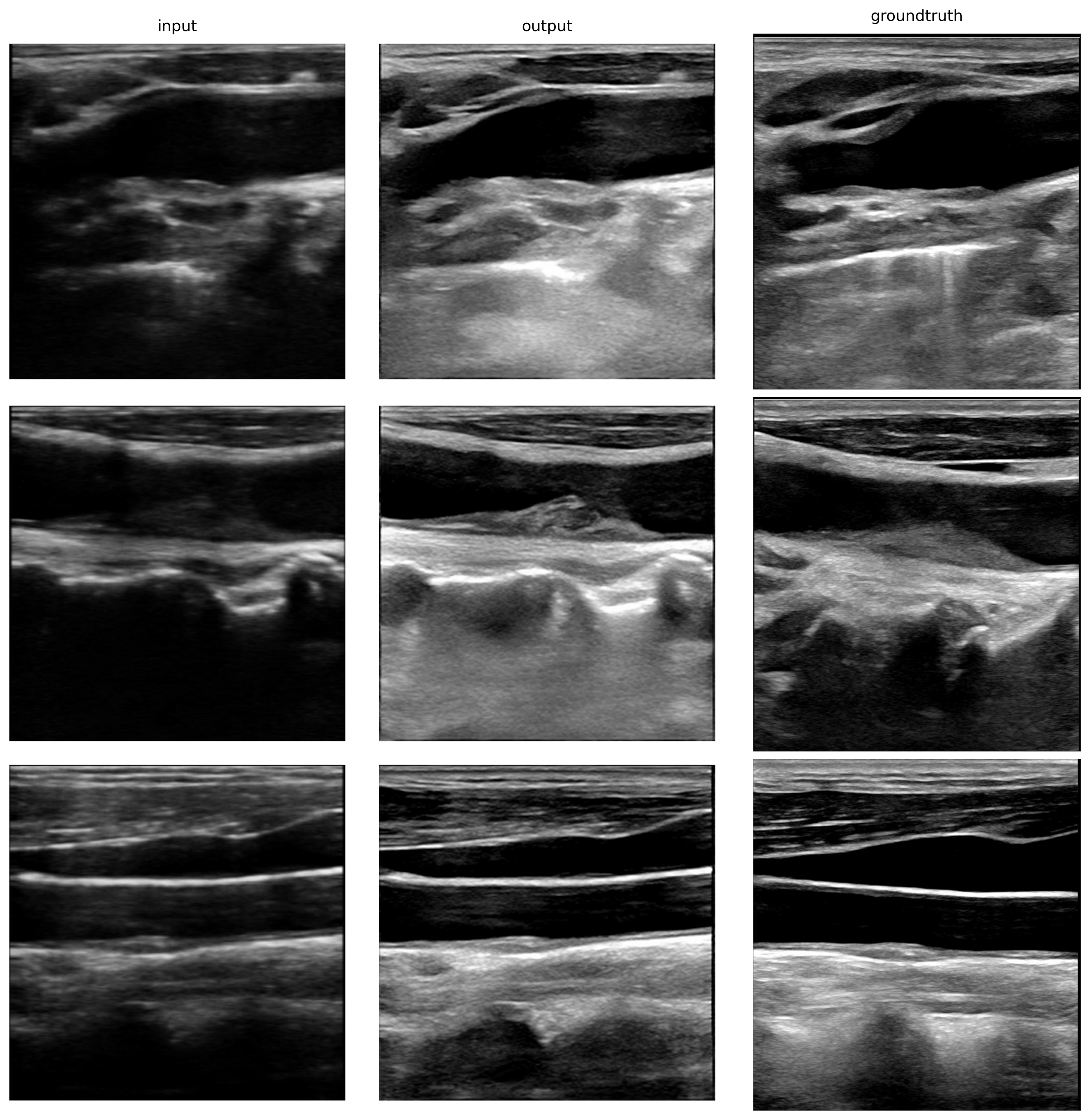
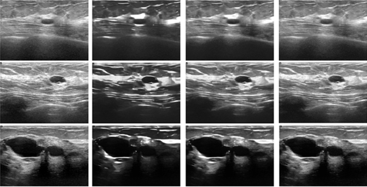
To overcome the challenges encountered in denoising ultrasound images, we propose a controllable method for denoising medical ultrasound images. This study mainly utilizes the TATLAB Toolbox to generate simulated noisy ultrasound image datasets and constructs an initial denoising model using the preprocessing of simulated noisy ultrasound images, the multi-level residual atrous spatial pyramid pooling (MRASPP) module, the nonlinear mapping convolutional neural network (NMCNNB) module, and the adaptive noise level and variable denoising intensity module. The model is trained through two rounds of inference.
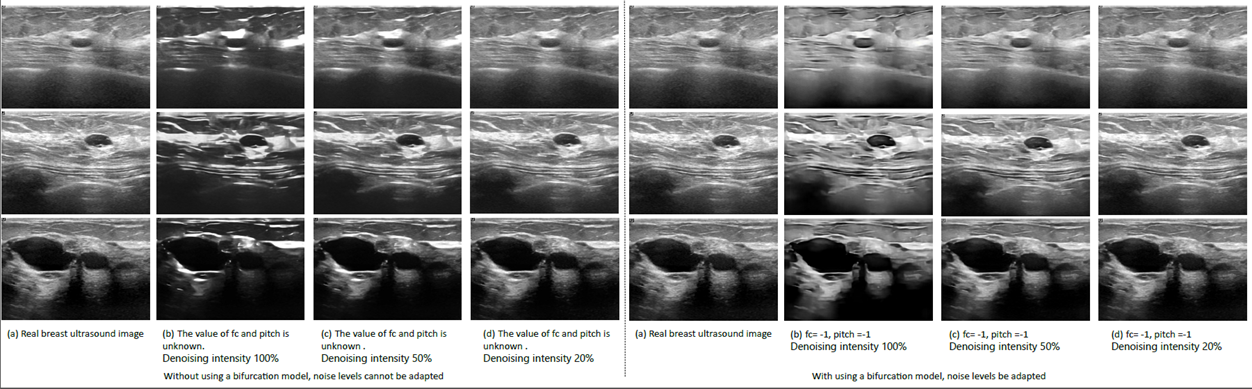
This method can adapt to a wider range of denoising scenarios, adaptively denoise the images, flexibly adjust the denoising intensity, effectively remove noise from medical ultrasound images, and preserve the details of the images. Compared to existing denoising methods, the average values of SSIM and PSNR have been improved by 1.67% and 1.28%, respectively. A comparison was made between the denoised and original images to evaluate their performance in downstream image classification tasks. After denoising real breast ultrasound images, the ACC and AUC of tumor benign-malignant classification tasks were improved by 1.09% and 2.83%, respectively.
The proposed controllable method for denoising medical ultrasound images, using two rounds of inference, can adaptively select the noise level parameter for effective denoising of ultrasound images formed by different ultrasound devices and settings. This method provides a new approach for denoising other types of medical images.
Please note that the translation and revision have been done to the best of my abilities, but there might still be room for improvement.
Project Example
Adaptive Dual-branch Network for Denoising and Artifact Removal in MR Images
Project Leaders
Mingfu Jiang
Partner Organisations
南京航天航空大學
Magnetic resonance (MR) images are essential for clinical diagnosis, yet their quality is often compromised by noise and artifacts, which can obscure anatomical details and reduce diagnostic accuracy. Existing deep learning approaches frequently address denoising and artifact removal as separate tasks, overlook pixel correlations and varying noise levels, require manual parameter tuning, and lack uncertainty evaluation, thereby limiting their practical utility in clinical settings.
This study aims to develop an Adaptive Dual-branch Network (ADbNet) for simultaneous denoising and artifact removal in MR images. The model is designed to adaptively handle varying noise and artifact levels, preserve complex structural details, and provide uncertainty estimates for the processed outputs to enhance reliability in downstream clinical tasks such as image segmentation.
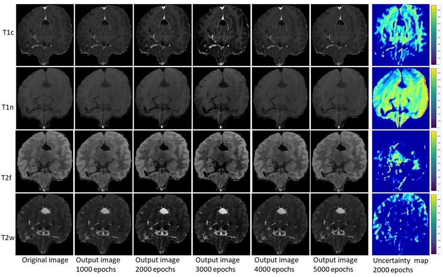
ADbNet integrates two specialized branches: a Heterogeneous Window Transformer Network (HWTNet) for adaptive denoising using both local and global feature modeling, and an improved U-Net based Artifact Removal Network (ARNet). Each branch is coupled with an estimation subnetwork—Noise Estimation Subnetwork (NES) and Artifact Estimation Subnetwork (AES)—to dynamically assess and adapt to input-specific degradation levels. The outputs of both branches are fused via an attention-based fusion module. Uncertainty is quantified during inference using Monte Carlo dropout. The model was trained and tested on 2000 brain MR scans from the Brats2021 dataset and 22 subject scans from the OpenNeuro dataset, incorporating synthetic Rician noise and real motion artifacts. Downstream segmentation performance was evaluated on the Medical Segmentation Decathlon (MSD) brain dataset (1666 cases) and a prostate MR dataset (203 examinations).
On the Brats2021 and OpenNeuro test sets, ADbNet achieved PSNR values of 27.23 ± 1.02 dB and 27.13 ± 1.32 dB, SSIM values of 0.9132 ± 0.0134 and 0.9121 ± 0.0458, and NMSE values of 0.0753 ± 0.0093 and 0.0765 ± 0.0091, respectively. For the downstream segmentation task, the denoised and artifact-corrected outputs led to average Dice score improvements of 2.27%, 1.33%, 1.37%, and 1.04% for T1n, T1c, T2f, and T2w MR modalities in the MSD brain dataset, and 1.11% and 1.18% for T2w and ADC sequences in the prostate dataset.
ADbNet effectively integrates adaptive denoising and artifact removal within a unified framework, significantly enhancing MR image quality and downstream segmentation accuracy. ADbNet explicitly accounts for spatially-varying noise characteristics and motion-induced artifacts commonly encountered in modern MRI systems. The incorporation of uncertainty estimation provides additional insight into result reliability, making the model a promising tool for clinical image enhancement and analysis. Future work will focus on computational optimization and extension to other anatomical regions.